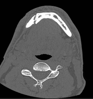
This right parasymphyseal mandibular fracture extends between a couple of teeth (no teeth lost--yet!):

 I also have a comminuted (multi-part) fracture of the left subcondylar mandible:
 Multiple fractures involving the maxillary sinuses: The alveolar ridge and hard palate were intact.
 Deviated nasal septum; blood in the maxillary sinuses:
 Small fractures of the anterior ethmoids, which extended into the frontal sinuses:
 These next couple of images are "coronal" images, oriented just as if I were looking at you. This one better shows the fractures of my ethmoid air cells (right across the bridge of my nose):
 Pterygoid plate fractures: There should be 2 nice, upside-down "V"-shaped bones near the middle of the image.


4 comments:
The 3d images are quite creepy
Jim - I have no idea what any of that stuff means, but we are so glad you are doing okay (you may disagree, I don't know).
Just wanted you to know we've been thinking about you and praying for you. Hang in there!
Elizabeth & Jason
James...
Did they at least make you more attractive during the surgery?
Steve
So... if I go into the hospital and tell them I want some 3D pictures of my skull, they can do that? Way cool.
Post a Comment